

1.3 Medical physics
Completion requirements

- Medical imaging refers to several different technologies that are used to view the human body in order to diagnose, monitor, or treat medical conditions.
- There are several imaging techniques that can provide imaging of biological samples.

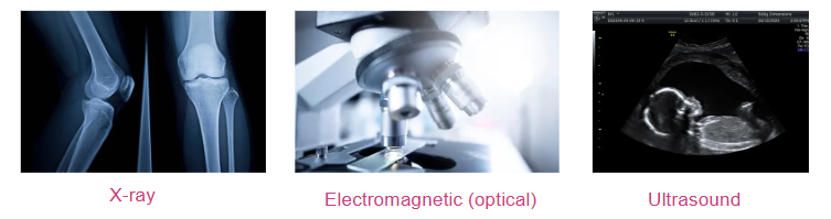

- An X-ray imaging is based on the absorption of X-rays as they pass through the different parts of a patient’s body.
- Differences in the densities of body tissue allow us to see inside the body by creating a shadow gram.





- This instrument consists of a small bell-shaped cavity attached to a hollow flexible tube.
- The bell is placed on the skin over the source of the body sound (such as the heart, intestines, or lungs).
- The sound is then conducted by the pipe to the ears of the examiner who evaluates the functioning of the organ.
- A stethoscope can detect sound waves with frequency ranging from tens to thousands of Hertz


- If the frequency of sound is higher than 20 KHz (0.02MHz), it is called ultrasonic or ultrasound.
- Typical frequencies used in medical ultrasound are 3.5-10 MHz.
- Ultrasonic waves penetrate tissue and are reflected, scattered and absorbed within it.
- The scattered and reflected ultrasound contains information about the form and structure of the tissue.

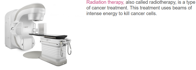
- The photons of X-rays and gamma-rays and the particles emitted by radioactive nuclei all have energies far greater than the energies that bind electrons to atoms and molecules.
- In controlled doses radiation can be used therapeutically. In the treatment of certain types of cancer.
- By careful placement of the radioactive material and by controlling the dose, the cancer cell can be destroyed without greatly damaging the healthy tissue.
Last modified: Wiixata, 14 Adoolessa 2025, 1:07 PM